参加活动:0 次 组织活动:0 次

最后登录2026-1-17
|
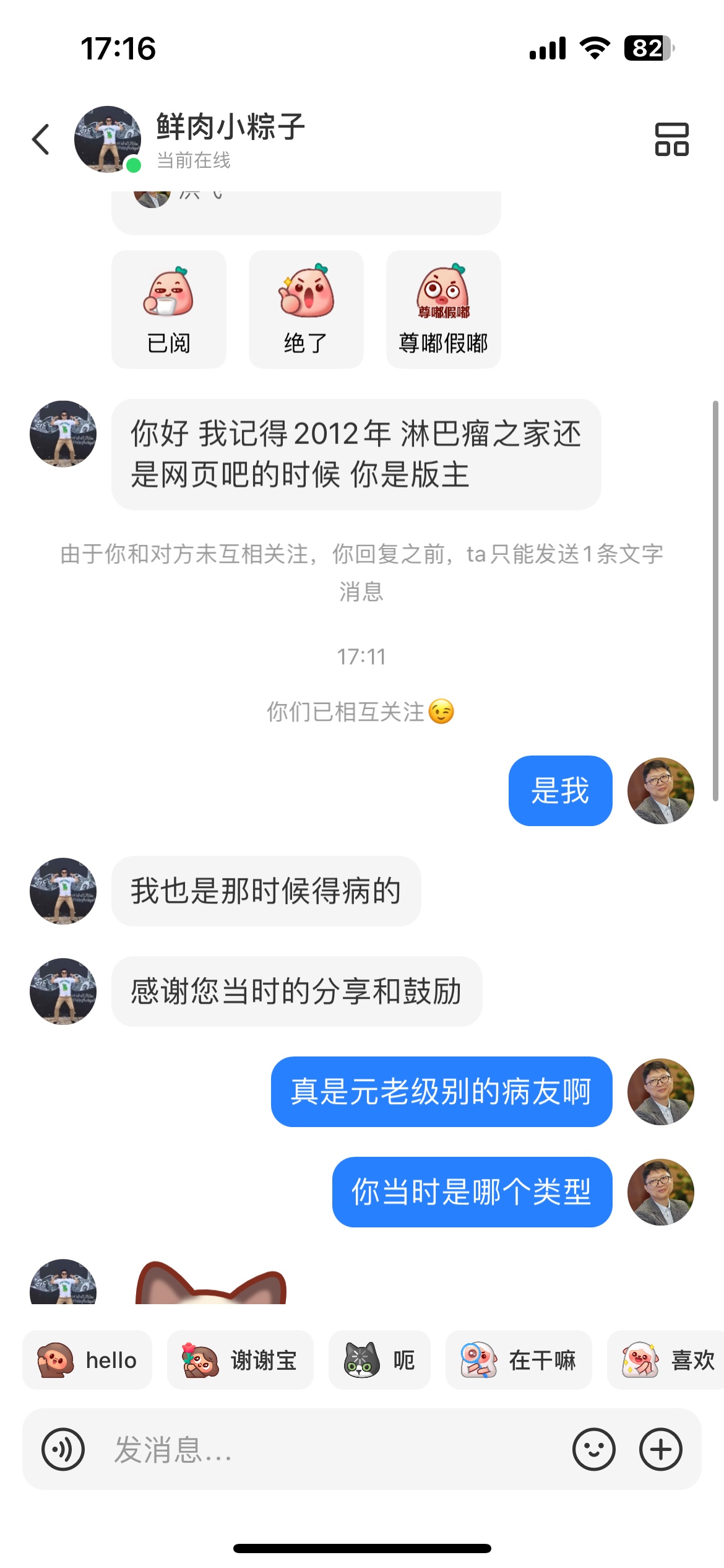
四疗后的petct结果可以看我原来的帖子,这是原来病灶都消了,但是中枢进展了吗?
我治疗前硬脑膜外有病灶,但没侵犯到中枢神经系统,每次也都有做腰穿,没有检测到淋巴瘤细胞,突然就好像中枢进展了。
Impression
A 2 to 3 mm tiny focus of intense tracer FDG uptake within the thecal sac of the very distal sacrum is new, and concerning for recurrent intrathecal metastatic disease, given presence of intracranial disease on prior MRI.
Previous noted heterogeneous uptake seen in the pelvis on prior PET/CT have resolved.
Intense uptake within the testicles bilaterally and uniformly is likely physiologic; recommend follow up by clinical exam.
Narrative
COMPARISON: PET/CT dated 2/10/2025. MRI dated 1/10/2025.
EXAM PURPOSE: Subsequent treatment planning.
ADDITIONAL HISTORY:
History of Burkitt lymphoma, follow-up, last therapy in March 2025.
FASTING BLOOD GLUCOSE: 104 mg/dl.
TECHNIQUE:
On 4/9/2025, the patient received intravenous injection of mCi 18-fluoro-2-deoxyglucose (FDG) for PET/CT imaging on a GE whole body PET CT scanner.
Imaging commenced after a 60 minute uptake.
CT TECHNIQUE:
Axial 1.5 mm CT with sagittal and coronal reformats performed after IV administration of non-ionic contrast of the head to thighs, without complications.
Otherwise, low resolution.
CT Dose: CTDi and DLP are available within the exam.
Vertex to mid thigh.
Multiplanar PET, CT, and PET/CT fused images were generated using iterative reconstruction and segmented attenuation correction techniques.
FINDINGS:
Typical Liver uptake, max SUV 3.8.
HEAD/NECK:
There is physiologic distribution of tracer in the pharynx, tonsils, salivary glands, and laryngeal regions.
No abnormal hypermetabolic activity is detected within the neck.
CHEST:
There is normal physiologic myocardial uptake of FDG. No hypermetabolic focus is identified in the chest.
ABDOMEN:
There is normal physiologic uptake of FDG in the liver, spleen, kidneys (with excretion into ureters and bladder), and gastrointestinal tract.
PELVIS:
There is physiologic excretion of FDG into the bladder. Intense uptake within the testicles bilaterally and uniformly is likely physiologic.
SKELETON:
There is a tiny 2 to 3 mm focus of intense tracer uptake, likely within the thecal sac of the very distal sacrum, SUV max of 5.0. This is new since prior exam.
The heterogeneous uptake seen in the pelvis on prior PET/CT has resolved.
No additional findings.
Geographic area of hypodensity within the central aspect of the liver is not FDG avid. This this is likely to represent fatty infiltration.
Continued dilatation of the pancreatic duct, but without obstructive lesion.
|
|
 /1
/1 


 窥视卡
窥视卡 雷达卡
雷达卡 发表于 2025-4-12 13:56:50
 提升卡
提升卡 置顶卡
置顶卡 沉默卡
沉默卡 喧嚣卡
喧嚣卡 变色卡
变色卡 抢沙发
抢沙发 显身卡
显身卡 楼主
楼主


 发表于 2025-4-12 17:19:35